(Please be aware that there will be some pretty gross pictures of hoof disections in this post - please try to check them out though, the knowledge gained is invaluable!)
 |
| An x-ray of a normal hoof. 'A' indicates P3. (B is the navicular bone, c is P2 or the short pastern bone, and D is P1 or the long pastern bone). |
 |
| A cross section of a cadaver leg. |
 |
| This is an awesome digital image of P3 in relation to the lateral cartilages (light blue) and tendons. |
Pedal Bone, Coffin Bone, P3, Distal Phalanx - this bone goes by a few different names. I like to refer to it as P3 as it is quicker to type and say!
P3 is the primary bone in the horse's hoof. To me, it looks like a little miniature version of a hoof - the outer hoof should match the bone on the inside. A bigger, flatter-soled P3 will show a flatter soled larger hoof. A very concave P3 bone will show as a hoof with magnificent concavity. There are soft tissues and blood vessels attached to P3 including the soft laminae that attach the bone to the hoof capsule.
 |
| Hoof with capsule removed showing the solar corium (the blood filled pad separating the sole and lower surface of P3), the soft laminae and the coronary corium from where the hoof horn grows. |
 |
| Look at the solar surface! The sole and frog corium (which is just a fancy name for soft tissues if you ask me) looks like a play-doh cast of the hoof! This shows why it is important to have sufficient sole depth. |
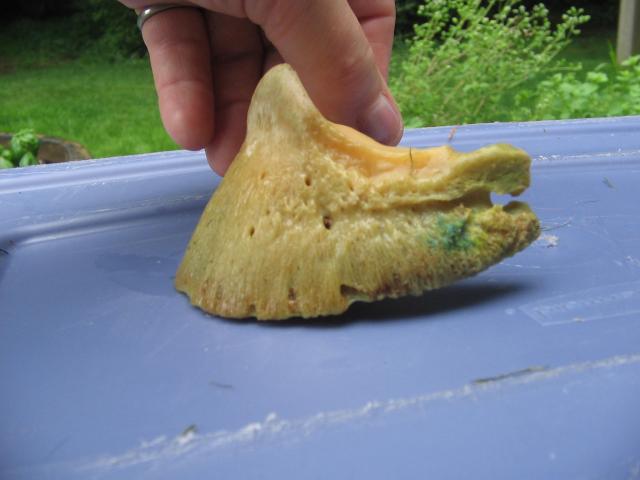
I did some googling and there are some VERY interesting pictures of P3 bones out there!
 |
| Various P3 bones of front feet from the top. The one top left has significant damage probably from an abscess or infection of some kind. |
 |
| Same bones as the picture above but viewed as if holding the hoof in your hand to be picked. Note the different soles and how they differ in concavity. |
 |
| As viewed from the side. |
 |
| Here is P3 in the hoof capsule |
 |
| And again on a shod hoof. Here it shows really well the concept that the collateral grooves (the valleys beside the frog) are a good indicator of sole depth - the shallower they are, the less sole you have. |
You will hear about P3 being talked about in relation to rotation and balance, particularly with cases of laminitis.
Anterior to posterior balance refers to the angle of P3 in relation to the ground surface. The accepted norm is 3-5 degrees rotation when viewed from the side. This is to allow some movement of P3 and the structures in the back half of the foot to 'sink' when the hoof is loaded and expands (that is why it is important to trim in quarter scoops to follow the live sole plane).
 |
| The red lines show the amount of rotation - imagine this hoof being loaded when taking a step - the angle made by the two red lines would close when loaded to absorb impact. |
Laminitis is as we all know when the laminitic structures loose their connection for a plethora of reasons and P3 can and often does rotate.
 |
| Here you can see the tip of P3 making the sole bulge just in front of the frog. Your brain should be screaming a warning at you if you ever pick up a hoof and see this! This is why a laminitic horse uses the infamous 'laminitis' stance - where a horse throws his hooves out in front of him and loads his heels to take the pressure off P3 and the excruciatingly painful laminae) |
 |
| An x-ray of a laminitic foot. See how P3 has rotated, the hoof wall has ripped away from the front surface of P3? See how the foot would bulge? |
The best way to see without an x-ray machine if the horse has an ideal or acceptable amount of rotation is to draw a line straight through the pastern and hoof - the angle of the hoof capsule should closely match that of the pastern.
 |
| Here is Gracie back in March this year - you can see the angle of the pastern vs the hoof is broken forward - the tip of P3 is is rotated slightly forward and down, which is evident in the flare on this hoof (most of which I removed). |
 |
| Here is Allie in April. The angle is broken slightly backwards - her P3 would be more ground parallel than Gracie's. Both are functioning, sound feet. |
What else would you add to this P3 post? :)


This is all good. Can't say I enjoy the cadaver photos but if you don't know how this all works, how do you know how to keep it working?
ReplyDeleteGood information and yes, ick, photos.
Thanks Barbara. This is why I put the warnings, as the cadaver pics can be quite graphic. I had a lot of trouble at first separating the fact I was looking at poor pony feet, but yes, how else do you learn? I'm still to do my own dissection, I have watched but can't cross that bridge to touching just yet.
ReplyDeleteGreat post! I was just trying to find your e-mail to respond to your message, but very informative! I did repsond to the paypal e-mail, so I know it went out. I want to keep it a secret so e=mail me at rehabdenali@yahoo.com and i'll reply! :)
ReplyDeleteThe last couple posts are quite good!
ReplyDeleteHello,
ReplyDeleteI´m writing a paper for school about horse´s hoof. Can I, please, use your picture "Here is P3 in the hoof capsule"? Of course, if you write me your full name and adress, I´m going to cite you :).
Regards,
Kristyna Hargitaiova, Czech republic
Kristyna, I don't own that photo anyway, I got it off Google Images. Thank you for asking though!!
ReplyDelete